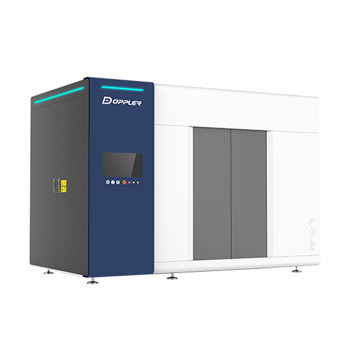



High-precision Inspection System for Castings and Forgings
Traditional NDT methods struggle to fully identify internal defects in castings and forgings due to their complex geometries and variable material properties. Conventional X-ray DR inspection requires multiple-angle scanning, which is time-consuming and likely to miss defects.
Developed by Doppler, the new cross-scale micron-level high-precision CT imaging system leverages the differential X-ray absorption of different materials in workpieces. It converts detector-captured attenuated X-ray signals into electrical signals, which are amplified, digitally converted and transmitted to a computer. By calculating the internal attenuation coefficient distribution, the system reconstructs images and generates high-precision 3D internal structure models of workpieces.
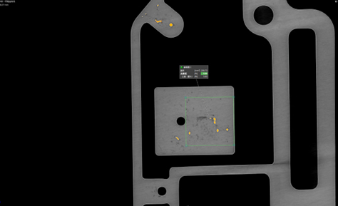
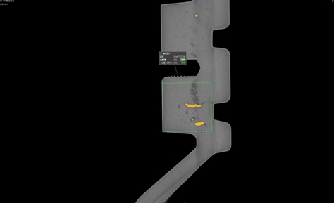
Breaking the constraints of traditional inspection, it significantly boosts testing efficiency and accuracy. Integrated with intelligent defect recognition technology, it clearly presents microscopic details, helping enterprises optimize production processes and improve overall manufacturing quality and efficiency.
Product Advantages
Intelligent Measurement
● Intelligent defect framing
● Visual defect measurement
Image Positioning
● Virtual sectioning, positioning and image segmentation
● Multi-dimensional image quality enhancement technology
Image Enhancement
● Multiple artifact correction technologies
● Image denoising and enhancement technologies
AI Algorithms
● Highly integrated AI reconstruction algorithms
● Automatic image feature recognition and measurement algorithms
An optional AI module with multiple network models is available to selectively improve image quality in a targeted manner.
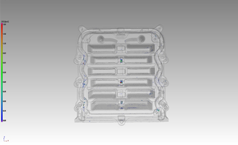
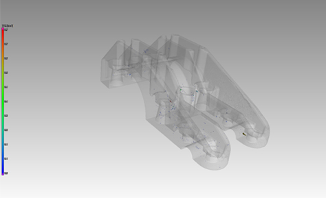
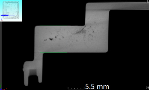
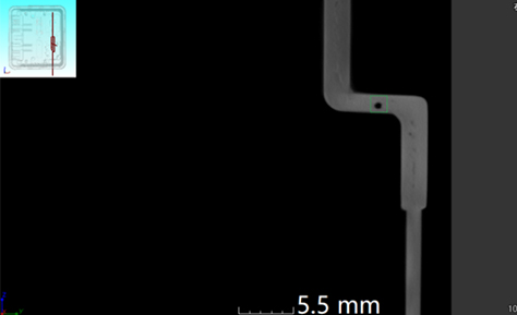
Testing Cases
Product Parameters

● Dual-ray source and dual-detector design with freely switchable micro-focus

● High-energy penetration: X-ray source voltage optional at 225KV/450KV, adapting to the thickness and density of various metal materials, castings and forgings


Item

Parameter

X-ray source focus

225KV:7μm

450KV:63μm

Detector pixels

Flat panel detector: 3072*3072

Linear array detector: 12 pixels/mm

Resolution (JIMA card test)

≤7μm

≤40μm

Pixel size

139 μm

(Flat panel detector)

Range

Diameter × Height: 280mm × 400mm (single scan)

Maximum imaging size with expanded field of view: Diameter × Height 400mm * 600mm

Exercise schedule

The sample stage can move in both the X and Y directions and rotate around the Z-axis, with a Y-axis travel ≥1600mm.

The X-ray source can move along the Z-axis, with a Z-axis travel ≥800mm.

The detector can move in three directions (X, Y, and Z), with an adjustable focal length (FDD) ≥1600mm and a Z-axis travel ≥800mm.

Sample stage load-bearing capacity

100kg

Radiation leakage rate

≤2.0μSV/h

Equipment dimensions (length*width*height)

5600*2700*3140(mm)

Equipment weight

40000 Kg


Back to List